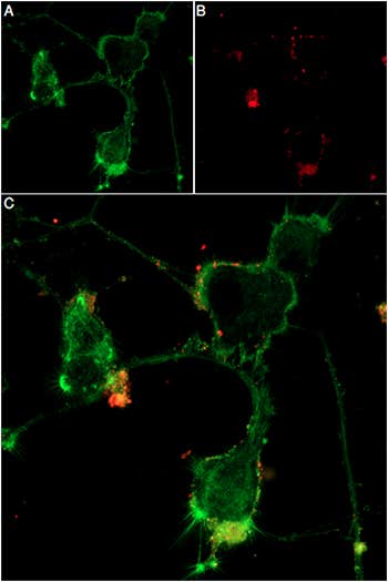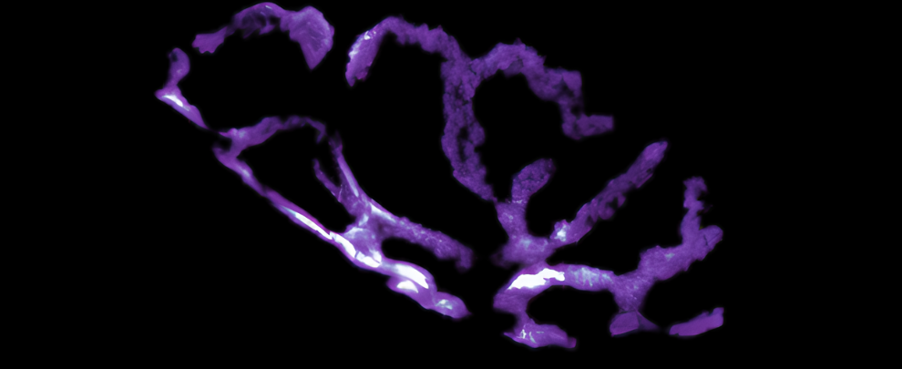
What if a tool borrowed from venomous snakes could give us a heap of new insight into a devastating disease? That’s exactly what α-bungarotoxin (α-BTX), a neurotoxin derived from snake venom that binds to nicotinic acetylcholine receptors (nAChRs), does in some fascinating amyotrophic lateral sclerosis (ALS) research. To really capitalize on the power of BTX, scientists employed a fluorescently labeled version of the toxin, transforming it into a versatile tool for studying the neuromuscular junction (NMJ).
In a study by Ionescu et al.,(1) α-BTX conjugated to fluorescein isothiocyanate (FITC) played a leading role. Why? Because it binds with high affinity and specificity to nAChRs – an essential part of nerve-to-muscle communication. And coupled with the conjugated FITC, the researchers we able to see the damage done to NMJs in a mouse model of ALS.
But the team didn’t stop at observing destruction. They went on to look at pridopidine, a compound with neuroprotective properties, to see if it could restore NMJ integrity. What they found was that under the fluorescent glow of our α-Bungarotoxin-FITC (#B-100-F), previously fragmented NMJs were brought back to life, with ~75% of junctions regaining structural integrity (Figure 1).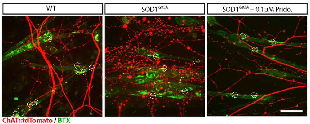
Figure 1. Representative images of in-vitro NMJs in the distal compartment display co-localized presynaptic motor axons (red) and AChR patches (green) on myocytes (circled by white dashed lines). The left panel shows that the co-localized axons in the WT co-cultures are intact, whereas axons in SOD1G93A co-cultures are degenerated (center panel). SOD1G93A co-cultures treated with 0.1 µM pridopidine display healthy NMJs and a reduced number of degenerated axons (right panel). Scale bar: 40 µm. Adapted from Ionesco et al. (2019) (1).
Over the course of several additional experiments, the researchers showed that ALS disrupts NMJs early in disease progression, even before motor neuron death, which supports the “dying-back” hypothesis. Their work also highlights the therapeutic potential of pridopidine, which restored NMJ integrity in ALS models – this means it could be a candidate to counteract mechanisms driving NMJ degeneration and slow disease progression.
Why Toxins?
So, why use a venom-derived toxin like α-BTX for this work? The answer lies in its targeting ability. α-BTX zeroes in on the extracellular domain of nAChRs, which allows researchers to confidently target this specific type of receptor. By conjugating it to FITC, the researchers here turned this snake toxin into a glowing marker to shed light on critical cellular events otherwise hidden in the biological maze.
What Else?
These ALS studies also highlight the power of combining neurotrophic support with precise molecular targeting. To promote neuronal health and function, the researchers employed Recombinant human BDNF protein (#B-250) and Recombinant human GDNF protein (#G-240). For enhanced detection sensitivity in quantification assays, they also used human BDNF-Biotin (#B-250-B), taking advantage of its compatibility with biotin-streptavidin systems.
What Does This Mean for Science?
The use of labeled toxins goes far beyond ALS research. These tools are usually passed over in favor of traditional antibodies. But the power and versatility of labelled toxins brings new options for research, allowing scientists to
- Map the architecture of complex systems, like synapses and receptor networks
- Investigate ion channel function and distribution simultaneously
- Track the real-time effects of drugs or experimental treatments
- Unravel dynamic interactions that underpin development and disease progression
For ALS, this approach helped uncover early pathological events, like NMJ disruption, to give scientists new targets for therapy. Beyond neurodegeneration, tools like this open avenues for probing diseases where cellular communication goes awry, from autoimmune disorders to cancer.
So, what’s next? As researchers refine these fluorescent probes and develop even more advanced labeling techniques, we’re set to gain even deeper insights into the hidden worlds of cellular biology. For now, α-BTX-FITC remains a glowing example of how venomous ingenuity is being transformed into a beacon of scientific discovery.
While FITC was the tool of choice in this study, Alomone Labs offers a complete panel of α-Bungarotoxin conjugates designed to meet diverse experimental needs. These include the native α-Bungarotoxin (#B-100), as well as fluorophore-conjugated variants such as ATTO Fluor-488 (#B-100-AG), ATTO Fluor-550 (#B-100-AY), ATTO Fluor-590 (#B-100-AR), ATTO Fluor-633 (#B-100-FR), and ATTO Fluor-647N (#B-100-FRN). For alternative detection strategies, α-Bungarotoxin-Biotin (#B-100-B) is also available. Each conjugate is produced and validated in-house to ensure exceptional performance across a wide range of bioassays.