Following NMDA receptor subtypes in live neurons
NMDA receptors are crucial for how neurons learn and adapt. They’re also complex beasts – built from multiple subunits, moving constantly across the neuronal membrane and changing roles as the brain develops.
To understand that behavior, you need to see what each subunit does. Not in fixed slices. Not averaged over a population. But in real time, on the surface of living cells. That’s exactly what researchers have been doing with a combination of cell surface-binding antibodies and quantum dots.
This is the third blog in a series on how cell surface-binding antibodies open up powerful experimental possibilities. And now we’re looking a little deeper, because to know what surface proteins do, you have to be able to watch them.
Real-Time Tracking of Membrane Proteins
Surface receptors don’t just sit there. They diffuse, cluster, anchor, un-anchor, and sometimes disappear entirely – and all of that affects how cells signal. But those dynamics don’t show up in fixed tissue. And intracellular antibodies won’t help – you can’t permeabilize a cell and expect it to behave like it’s alive. That’s why researchers turn to antibodies that bind cell-surface epitopes and then tag them with quantum dots.
Quantum dots are tiny semiconductor particles that glow brightly and don’t bleach over time. Attach them to an extracellular antibody, and you can track individual receptors on the surface of a live cell for minutes at a time, with nanometer precision.
It’s a method that’s opened up a whole new view of membrane biology. And a series of papers over the last few years has shown just how powerful it is.
The Setup: Quantum Dots Meet Subtype-Specific Antibodies
Each of these studies starts with the same basic strategy:
- Use a subtype-specific extracellular antibody (from Alomone Labs) to target either GluN2A or GluN2B, two key NMDA receptor subunits
- Apply a quantum dot-conjugated secondary antibody to tag the receptor
- Track single receptors as they move across the membrane of live hippocampal neurons
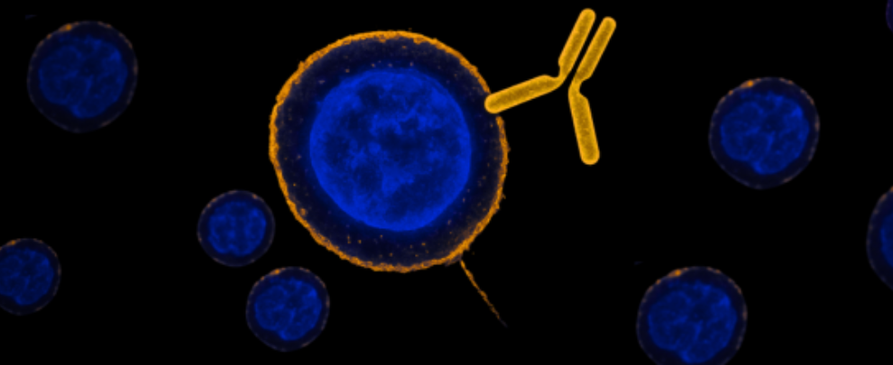
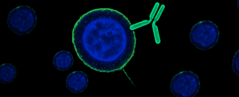
This only works if the primary antibody binds a truly extracellular epitope – and does so without disrupting receptor behavior. Our Anti-GluN2A (extracellular) antibody (#AGC-002) and Anti-GluN2B (extracellular) antibody (#AGC-003) (Figure 1) antibodies have been used extensively in this setup. Both bind to external domains and preserve receptor function, which makes them ideal for live-cell tracking.
In this experiment, for example, the researchers used advanced super-resolution microscopy techniques, including dSTORM analysis, to investigate how GluN2A and GluN2B NMDA receptor subtypes are organized at the nanoscale on the surface of live neurons. Live-cell labeling was performed using our extracellular antibodies against GluN2A and GluN2B, enabling specific detection of surface-expressed receptors. By comparing conventional epifluorescence with high-resolution imaging, they visualized and quantified differences in cluster size, molecular density, and nanodomain organization – revealing distinct structural patterns between the two receptor subtypes and shedding light on their differential roles in synaptic maturation.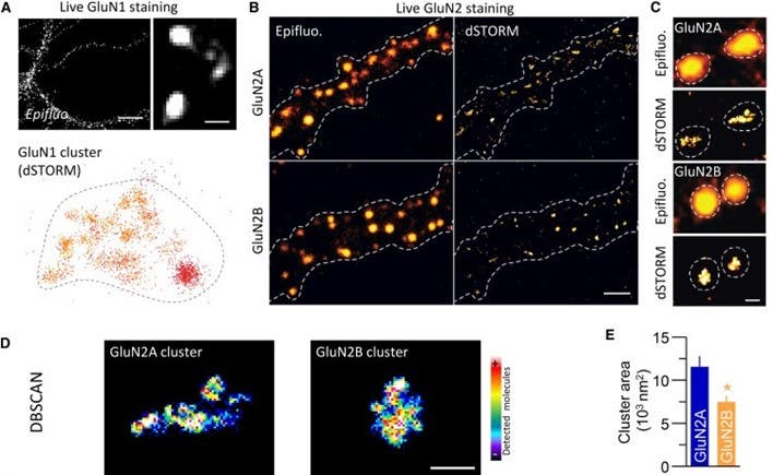
Figure 1. Super-resolution imaging reveals differences in the nanoscale organization of GluN2A and GluN2B NMDA receptors in neurons
(Adapted and cropped from Kellermayer et al., 2018, Neuron. https://www.sciencedirect.com/science/article/pii/S0896627318307852). A) Visualization of surface GluN1 subunits (a shared component of NMDA receptors) using epifluorescence (top left) and super-resolution dSTORM microscopy (top right). Bottom: dSTORM image showing the fine-grained structure of a GluN1 receptor cluster. Dashed outline marks the border of the cluster. Scale bar = 200 nm. B) Surface staining of GluN2A and GluN2B subunits on live neurons shows receptor distribution along dendrites. Images compare standard epifluorescence (left) and high-resolution dSTORM imaging (right). Dashed lines outline dendritic spines. Scale bar = 1 μm. C) Higher-magnification comparison of single GluN2A and GluN2B clusters imaged by epifluorescence (top) and dSTORM (bottom) reveals that GluN2B clusters tend to appear more compact and defined in super-resolution imaging. Scale bar = 200 nm. D) DBSCAN-based analysis of single clusters highlights the molecular density within GluN2A and GluN2B clusters. Warmer colors represent regions with more detected receptor molecules. Scale bar = 200 nm. E) Quantification shows that GluN2A clusters are significantly larger than GluN2B clusters.
What the Tracking Reveals
D-serine rapidly increases GluN2B mobility – but not GluN2A
When researchers added D-serine, a natural co-agonist of NMDA receptors, GluN2B-containing receptors became more mobile and less anchored at the synapse. GluN2A stayed stable. That difference only became clear thanks to live quantum dot tracking using the subtype-specific extracellular antibodies (1).
GluN2A and GluN2B cluster in distinct nanodomains
Super-resolution imaging combined with quantum dot tracking showed that GluN2A and GluN2B occupy different zones within the synapse. These zones can rearrange in response to signaling – and that rearrangement tunes synaptic strength, even without changing receptor numbers (2).
The proteasome selectively increases GluN2B mobility
Activating the synaptic proteasome increases GluN2B lateral diffusion – but has no effect on GluN2A. Knock down GluN2B, and the proteasome itself disperses. Again, these reciprocal dynamics only show up when you track each receptor subtype live (1).
GluN3A reshapes GluN2A surface dynamics during development
When GluN3A is expressed early in development, it selectively destabilizes GluN2A, making it more mobile and less synaptically anchored. GluN2B is unaffected. This finding helps explain how NMDAR composition changes as networks mature (3).
Why this Method Works
These results aren’t just about NMDA receptors. They’re about what you can do with the right antibody.
This method works because the antibodies:
- Bind live, non-permeabilized cells
- Recognize external protein domains with high specificity
- Preserve the natural behavior of the receptor
- Are compatible with real-time tracking systems like quantum dots
Once this setup is in place, you’re free to follow surface receptors at the single-molecule level. You can see how co-agonists, signaling proteins, or developmental cues shift their behavior – and do it in real time.
In short, if your receptor is on the surface, and your question is about when, where, or how it moves, you can use this method. And you can build it using antibodies that are already validated for this exact application.
Want to study trafficking? Subtype segregation? Activity-dependent anchoring? Start with the cell-surface antibody. The quantum dots come next.
Up Next in the Series
In the final part of our series on cell surface-binding antibodies, we’ll take a closer look at how these antibodies are being used in vivo.
Missed the Other Parts? Catch Up Here
- Part 1: Target the surface, stay out of the cell
• How cell surface-binding antibodies reveal real-time ion channel trafficking - Part 2: Aim, bind, deliver – with light
• Non-lethal targeted cell delivery using gold nanoparticles and laser pulses
 (extracellular) Antibody")
 (extracellular) Antibody")