Multiplexing with complementary labels
So far, we have looked at live tracking, targeted delivery, quantum dot dynamics, and in vivo biology. But there is another tool in the extracellular antibody set we have not covered: multiplex flow cytometry.
By using antibodies conjugated to distinct fluorophores, such as allophycocyanin (APC), fluorescein isothiocyanate (FITC), or phycoerythrin (PE), you can detect multiple targets on the same cells, at the same time, in the same tube. This approach delivers more data per run, reduces sample use, and increases biological insight.
From One Color to Many – Why Multiplexing Matters
Directly conjugated antibodies make flow cytometry faster and simpler – no need for secondary antibodies, and less background noise. Here are three different panels from a recent experiment showing how you can use our APC- and PE-conjugated antibodies to dig deeper into immune and metabolic biology. All antibodies come with matched isotype controls to help you validate your data with confidence.
Defining Microglia in the Mouse Brain
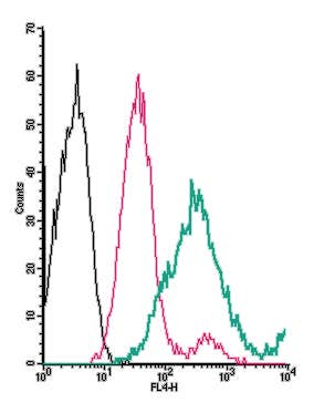
For example, combining Anti-TMEM119 (extracellular)-APC antibody (#ANR-175-APC) with Anti-P2Y12 Receptor (extracellular)-PE antibody (#APR-020-PE) (Figure 1), helps clearly identify microglia in mouse brain tissue.
TMEM119 and P2Y12 are two of the most trusted markers for homeostatic microglia. TMEM119 is found only in microglia (not infiltrating macrophages), while P2Y12 is involved in how microglia sense and respond to injury in the brain. Both proteins sit on the cell surface, making them easy targets for flow with extracellular antibodies. This combo supports refined understanding of neuroimmune responses in various mouse brain models.
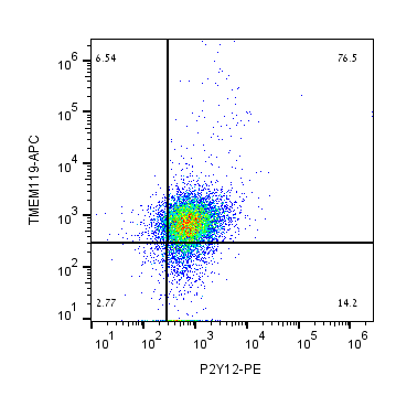
Figure 1. Identification of microglia subtypes in mouse brain using multiplex flow cytometry. Mouse microglia stained with Anti-TMEM119 (extracellular)-APC Antibody (#ANR-175-APC) and Anti-P2Y12 Receptor (extracellular)-PE Antibody (#APR-020-PE). These two surface markers help clearly identify microglia in the brain.
Looking at Metabolic Transporters in T Cells
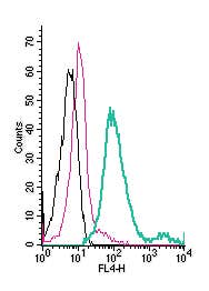
Pairing Anti-MCT1 (SLC16A1) (extracellular)-APC antibody (#AMT-011-APC) with Anti-Human ASCT2/SLC1A5 (extracellular)-PE antibody (#ANT-083-PE) in Jurkat T-cells (Figure 2) allows simultaneous assessment of two key transporters in metabolic studies.
Why these two? Because MCT1 and ASCT2 are key transporters involved in how T cells take up nutrients. MCT1 moves lactate in and out of cells (important when cells are highly active), and ASCT2 brings in glutamine, a fuel for growth and division. Together, they give you a picture of how the cell is handling energy and metabolism –relevant in immunometabolism studies, where metabolism and immune function go hand in hand.
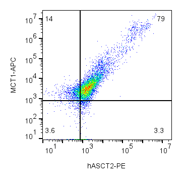
Figure 2. Metabolic transporter profiling in human T cells using multiplex flow cytometry. Human Jurkat T-cells stained with Anti-MCT1 (SLC16A1) (extracellular)-APC (#AMT-011-APC) and Anti-Human ASCT2/SLC1A5 (extracellular)-PE (#ANT-083-PE), two key transporters involved in immune cell metabolism.
Adrenergic Receptors in Macrophages
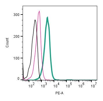
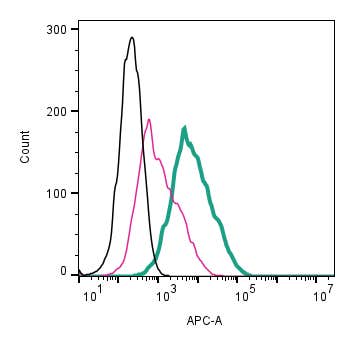
For this one, we looked at mouse J774 macrophages stained with Anti-β1-Adrenergic Receptor (extracellular)-APC antibody (#AAR-023-APC) and Anti-β2-Adrenergic Receptor (extracellular)-PE antibody (#AAR-016-PE) (Figure 3).
These two receptors – β1 and β2 – are how macrophages “listen” to signals from the nervous system. They’re part of a the GPCR group and they help regulate inflammation, migration, and even tissue repair. If you’re studying how stress, nerves, or inflammation affect immune cells, this panel is a great way to track those changes directly.
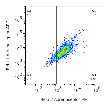
Figure 3. Adrenergic receptor expression in mouse macrophages revealed by multiplex flow cytometry. Mouse J774 macrophages stained with Anti-β1-Adrenergic Receptor (extracellular)-APC Antibody (#AAR-023-APC) and Anti-β2-Adrenergic Receptor (extracellular)-PE Antibody (#AAR-016-PE). These surface GPCRs respond to stress hormones and play important roles in immune regulation, particularly by influencing inflammation and macrophage activity.
More Conjugates, More Flexibility
One of the nice things about using surface-binding antibodies for flow cytometry is how flexible the setup can be. Most of our extracellular antibodies are available in a range of fluorophore conjugates – not just APC and PE, but also FITC, Atto Fluor® 488-647, and a mFluor Violet™ 450 that works well on many standard cytometers. That means you can mix and match depending on what lasers and filters your instrument supports, or how many channels you’re trying to squeeze into a single run. You can check out all available formats for each product on the extracellular antibody page.
Tools to Help You Plan Your Panel
If you’re building a flow panel with multiple colors, choosing the right antibodies is only part of the job. You also need to make sure the fluorophores don’t overlap too much, and that your cytometer can detect them cleanly. To make that easier, we’ve put together two simple tools: the Panel Builder lets you pick and preview antibody-fluor combinations based on your targets, and the Spectra Viewer helps you check for emission overlap before you hit the lab. They’re especially handy if you’re working with limited cell numbers or trying to streamline your staining workflow.
With careful panel design and proper compensation, researchers can run four to six markers simultaneously on standard cytometers, and even more on spectral or mass systems. This increases data richness without adding technical complexity.
The Payoff of Multiplex Flow Cytometry
With the right set of extracellular antibodies, researchers can extract more from each sample. Multiplex flow cytometry uncovers complex relationships, co-expression patterns, rare states and biological shifts that single-marker experiments miss. And because these antibodies bind external epitopes, the measured signal reflects biology, not processing artefacts.