Directly conjugated primary antibodies reduce background, save time, and simplify multiplex assays in imaging and flow cytometry. We now offer a fully validated portfolio of conjugated antibodies – all made and tested in house – that target key ion channels, receptors, and markers, and span the violet to far-red light spectrum.
Why Use Directly Conjugated Antibodies?
Conjugating the fluorophore directly to the primary antibody removes the need for a secondary detection step. This reduces nonspecific background, shortens workflows, and increases multiplexing flexibility – critical when combining multiple markers or working with delicate samples in immunohistochemistry (IHC), immunocytochemistry (ICC), or flow cytometry.
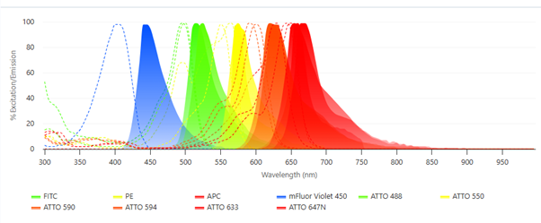
Fluorophore Coverage: Violet to Far-Red
You can now choose antibodies – and toxins – conjugated to fluorophores that span the spectrum, from ~445 nm (violet) to ~670 nm (far-red), allowing you to run multi-color experiments without worrying about spectral overlap.
New ATTO Fluor 647N-conjugated Antibodies and Toxin
These include the following:
- Anti-GFAP-ATTO Fluor-647N Antibody (#AFP-001-FRN) – mouse and rat-specific; validated for multicolor IHC and ICC.
- Anti-TRPV1 (VR1)-ATTO Fluor-647N Antibody (#ACC-030-FRN) – mouse, rat, and human-specific; validated for multicolor IHC and ICC.
- Anti-KCNQ1 (extracellular)- ATTO Fluor-647N Antibody (#APC-168-FRN) – a cell surface-binding antibody, mouse, rat, and human specific; validated for live cell imaging and direct flow cytometry.
- DkTx-ATTO 647N (#STD-010-FRN) – a synthetic conjugated toxin for TRPV1 research, useful in advanced functional and localization studies as well as live cell imaging; bioassayed in rat dorsal root ganglion cells and frozen sections, mouse BV-2 microglia cells, and Xenopus.
ATTO 647N is a high-performance far-red dye (Ex ~645 nm, Em ~670 nm) with excellent photostability and quantum yield. It’s especially suited to super-resolution microscopy, such as SIM and STED, and can offer high contrast even in autofluorescent tissues.
New mFluor™ Violet 450-conjugated Antibodies
These are all a cell surface-binding antibodies, mouse, rat, and human specific, and validated for live cell imaging and direct flow cytometry. They include:
- Anti-P2Y12 Receptor (extracellular)-mFluor™ Violet 450 Antibody (#APR-020-V)
- Anti-β1-Adrenergic Receptor (extracellular)-mFluor™ Violet 450 Antibody (#AAR-023-V)
- Anti-xCT/SLC7A11 (extracellular)-mFluor™ Violet 450 Antibody (#ANT-111-V)
mFluor Violet 450 (Ex ~405 nm, Em ~445 nm) is designed for flow cytometry using the 405 nm laser line and fits standard 450/50 detectors. It serves as a direct replacement for Pacific Blue™ or Brilliant Violet™ 421 dyes, for multi-parameter flow cytometry with minimal spillover.
Application Highlights
Immunohistochemistry and Immunocytochemistry
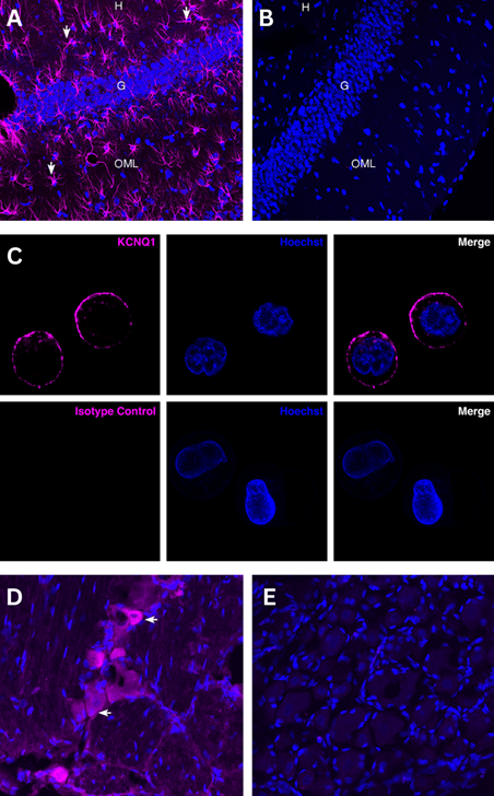
Figure 1. Immunohistochemistry and immunocytochemistry staining using conjugated antibodies. Top panel: Immunohistochemical staining of perfusion-fixed frozen rat brain sections with Anti-GFAP-ATTO Fluor-647N Antibody (#AFP-001-FRN), (1:80). A) Staining in the hippocampal dentate gyrus region, shows GFAP immunoreactivity (purple) in astrocytes (arrows). B) Staining of sequential sections with Rabbit IgG Isotype Control-ATTO Fluor-647N (#RIC-001-FRN), (1:80), demonstrate the low background signal. Cell nuclei are stained with DAPI (blue). H = hilus, G = granule layer, OML = outer molecular layer. Middle panel: C) Detection of KCNQ1 in Human COLO-205 Cells KCNQ1 was detected in live COLO-205 colon adenocarcinoma cells by confocal microscopy using Anti-KCNQ1 (extracellular)-ATTO Fluor-647N Antibody (#APC-168-FRN), (1:25, magenta). Nuclei were stained with Hoechst 33342 (blue). Control staining was performed using Rabbit IgG Isotype Control-ATTO Fluor-647N (#RIC-001-FRN), (1:25). Bottom panel: TRPV1 in rat dorsal root ganglion (DRG) Frozen DRG sections from a paraformaldehyde-perfused rat were incubated with Anti-TRPV1 (VR1)-ATTO Fluor-647N Antibody (#ACC-030-FRN), (1:100, magenta). D) TRPV1 immunoreactivity was observed in some DRG cells (arrows). E) Pre-incubation of the primary antibody with the TRPV1/VR1 Blocking Peptide (#BLP-CC030) suppressed the staining. Nuclear staining with DAPI (blue).
Flow Cytometry and Live Cell Imaging
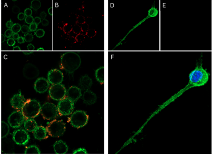
Figure 2. Live cell imaging with a fluorescently labeled toxin. Left panel: Live cell imaging of DkTx-ATTO Fluor-647N in live intact mouse BV-2 microglia cells. A) Plasma membranes were stained with CellMask™ Actin 1X solution (green). B) The same cells were then labeled with DkTx-ATTO Fluor-647N (#STD-010-FRN), (0.1 µM, red). C) Overlay image showing the distribution of DkTx across BV-2 cells. Right panel: Competition assay with unlabeled DkTx in rat dorsal root ganglion (DRG) neurons. D) DRG neurons cultured in DMEM/F12 supplemented with 100 ng/ml native mouse NGF 2.5S (99%) (#N-240) for 5 days, stained with CellMask™ Actin 1X solution (green). E) Pre-incubation with unlabeled DkTx (#STD-010), (100 µM) followed by staining with DkTx-ATTO Fluor-647N (0.1 µM) abolishes fluorescent signal. F) Absence of ATTO Fluor-647N staining (red) confirms specific competition by unlabeled DkTx. Nuclei were counterstained with Hoechst 33342 (blue).
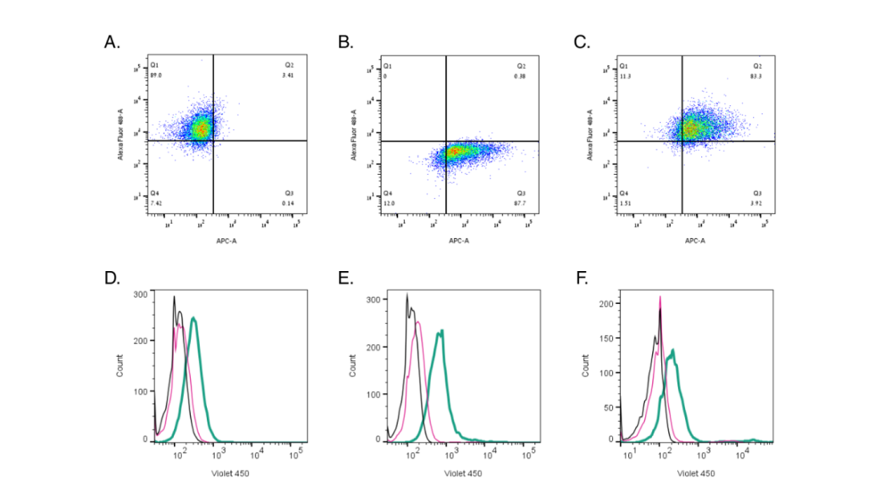
Figure 3. Flow cytometry using conjugated antibodies and a toxin. Top panel: Direct flow cytometry of DkTx in live intact mouse BV-2 microglia cells. A) BV-2 cells + Anti-TRPV1 (VR1) (extracellular) Antibody (#ACC-029) (1:100) + 2 µg/ml Goat anti-Rabbit IgG Secondary Antibody, Alexa Fluor™ 488. B) BV-2 cells + 100 nM DkTx-ATTO Fluor-647N (#STD-010-FRN). C) BV-2 cells + Anti-TRPV1 (VR1) (extracellular) Antibody (1:100) + 2 µg/ml Goat anti-Rabbit IgG Secondary Antibody, Alexa Fluor™ 488 + 100 nM DkTx-ATTO Fluor-647N. D) Cell surface detection of P2RY12 by direct flow cytometry in live intact mouse SIM-A9 microglia cells: Black) Cells. Red) Cells + Rabbit IgG isotype control-mFluor™ Violet 450 (#RIC-001-V). Green) Cells + Anti-P2Y12 Receptor (extracellular)-mFluor™ Violet 450 Antibody (#APR-020-V), (2.5µg). E) Cell surface detection of β1-Adrenergic Receptor by direct flow cytometry in live intact mouse J774 macrophage cells: Black) Cells. Red) Cells + Rabbit IgG isotype control- mFluor™ Violet 450 (#RIC-001-V). Green) Cells + Anti-β1-Adrenergic Receptor (extracellular)- mFluor™ Violet 450 Antibody (#AAR-023-V), (2.5µg). F) Cell surface detection of xCT/SLC7A11 by direct flow cytometry in live intact human U-87 MG glioblastoma cells: Black) Cells. Red) Cells + Rabbit IgG isotype control- mFluor™ Violet 450 (#RIC-001-V). Green) Cells + Anti-xCT/SLC7A11 (extracellular)- mFluor™ Violet 450 Antibody (#ANT-111-V), (2.5µg).
Why ATTO 647N and mFluor Violet 450?
We tend to use ATTO fluorophores for their photostability and low background. Compared to traditional labels like FITC or Alexa Fluor, ATTO dyes resist photobleaching during extended imaging sessions (1). ATTO 647N offers strong absorption, high photostability, and minimal sensitivity to atmospheric ozone, which make it ideal for demanding imaging applications, including single-molecule detection and super-resolution microscopy. At the other end of the spectrum, mFluor Violet 450 brings reliable brightness and compatibility with existing flow cytometry setups, without the compensation complexity often associated with tandem dyes.
Practical Tips
- Confirm antibody specificity using knockout controls or blocking peptides.
- Check the specific validation data found on all of data sheets for application consistency.
- For specialized imaging systems, get in touch with our Scientific Support team to help match fluorophores to your exact laser and detector setup.
- It’s always worth considering there will be reduced signal amplification with directly conjugated antibodies compared to indirect methods, since the secondary antibodies amplify your signal. This isn’t an error but a known trade-off you should be aware of.
| Antibody | Target | Applications | Fluorophore | Excitation (nm) | Emission (nm) | Spectrum Pick |
| AFP-001-FRN | Glial fibrillary acidic protein (GFAP) | IHC, ICC | ATTO 647N | ~645 | ~670 | Far-red |
| ACC-030-FRN | Transient receptor potential cation channel subfamily V member 1 (TRPV1) | IHC, ICC | ATTO 647N | ~645 | ~670 | Far-red |
| APC-168-FRN | KV7.1, Voltage-gated potassium channel QKT subfamily member 1 | Flow, live cell imaging | ATTO 647N | ~645 | ~670 | Far-red |
| STD-010-FRN | Tau-theraphotoxin-Hs1a-K2, targeting TRPV1 | Advanced functional studies, live cell imaging | ATTO 647N | ~645 | ~670 | Far-red |
| APR-020-V | P2Y purinoceptor 12 (P2RY12) | Flow, live cell imaging | mFluor Violet 450* | ~405 | ~445 | Violet |
| AAR-023-V | Beta-1 adrenoceptor | Flow, live cell imaging | mFluor Violet 450* | ~405 | ~445 | Violet |
| ANT-111-V | Cystine/glutamate transporter | Flow, live cell imaging | mFluor Violet 450* | ~405 | ~445 | Violet |
* When using an antibody conjugated to the mFluor™ Violet 450 in flow cytometers, it is best detected using the 405 nm laser lane and a filter set of 450/50.
Cover the Whole Spectrum
With ready-to-use antibodies conjugated to high-performance fluorophores, Alomone Labs gives you the reagents and confidence to design multiplexed, low-background experiments across imaging and flow cytometry platforms, from violet to far-red.
Custom conjugation services are also available so you can get exactly what your experiments need.
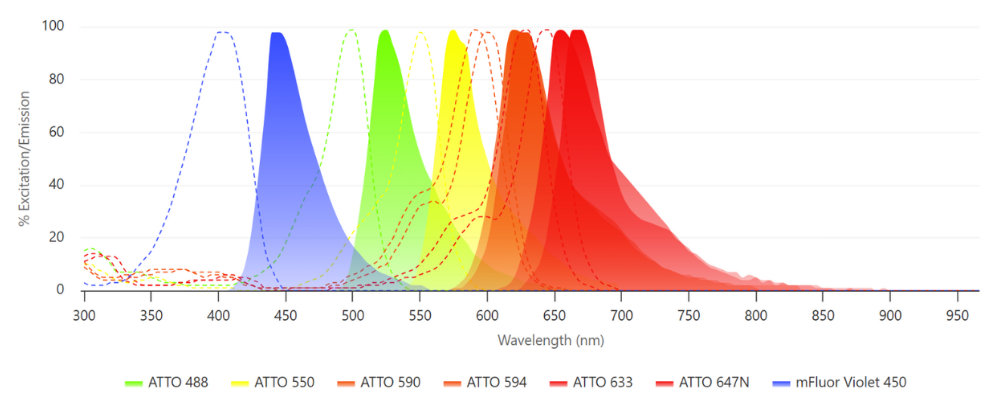
If you need to compare fluorophore spectra or check for potential overlap, our Spectra Viewer gives you everything in one place. Plot excitation and emission curves, adjust filters, and see where things might clash. It’s a clear, visual way to plan smarter panels and avoid compensation issues later on.
If you want to go straight to picking out antibodies plus the conjugated fluorophore, head over to the Panel Builder. This will help pick out the perfect antibody combination for assays like flow cytometry or just multiplex options. Simply choose your targets, pick your antibodies, and it maps out the best possible fluorophore combinations for your setup. Less guesswork, cleaner data.


-ATTO Fluor-647N Antibody")
-ATTO Fluor-647N Antibody")

-mFluor™ Violet 450 Antibody")

")

 (extracellular) Antibody")