Microglial dysfunction is a factor in neurological disorders such as schizophrenia and Alzheimer’s disease (AD). A better understanding of any pathological contribution from microglia requires a model that mimics what you observe in humans. Currently, these models may originate from human induced pluripotent stem cell (hiPSC)-derived microglia. However, differentiating hiPSCs into microglia is challenging and often slow, making them unsuitable for high throughput clinical trials. There’s also the issue that induced microglia (iMG) don’t always behave like microglia in vivo.
However, new research has generated iMGs from monocytes and validated these immunologically, behaviorally, and genetically (1). The iMGs were generated in under 2 weeks; a significant reduction from the 40 days it often takes to derive microglia from hiPSCs. The resulting iMGs could have real potential in understanding neurological diseases, such as schizophrenia and AD.
A Quick Introduction to Microglia
Microglia – the immune cells of the central nervous system – are essential for normal brain functions. When everything is working as it should, microglia play a role in neurogenesis, synaptic pruning, and phagocytosis when not participating in immune surveillance. However, when stressed or overstimulated, microglia can become activated and shift to their proinflammatory state (M1). Hyperactive microglia release neurotoxic factors and cytokines [interleukin (IL)-1β, IL-6, as well as TNF-α] and, if activated for too long, can induce neuronal apoptosis and brain damage observed in several neurological (2) and neurodegenerative disorders (3).
Microglia That Look Like Microglia
The first step in validating these iMGs was assessing their morphology. Under phase-contrast, iMGs started branching at day 5, and by day 10–14 they resembled typical resting microglia, with a small cell soma and multiple branches. Induced macrophages (iMacs) also had the typical “fried-egg” shape with enlarged circular cell bodies.
The investigators then focused their attention on microglia-specific markers, such as transmembrane protein 119 (TMEM119) and purinergic receptor P2RY12. These are unique microglia markers that are absent from macrophages. Using anti-TMEM119 antibody and Alomone’s Anti-P2Y12 Receptor (extracellular) Antibody (#APR-020), it was clear that the TMEM119 and P2RY12 markers were expressed by iMG cells but were absent from iMacs (Figure 1) as expected. This nicely illustrated how TMEM119 and P2RY12 can be used to distinguish iMG cells from monocyte-derived macrophages.
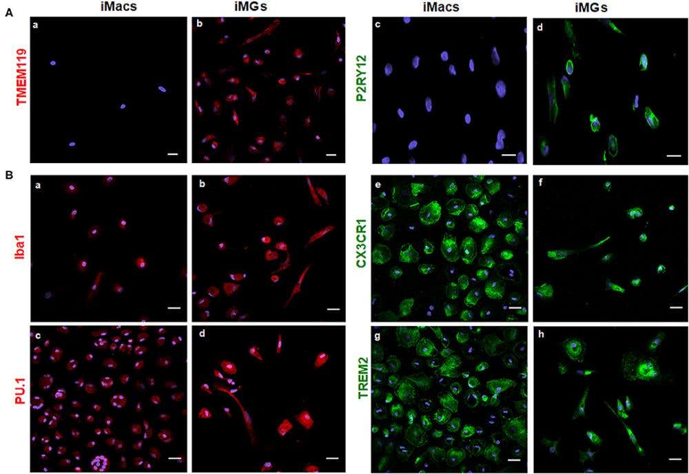
Figure 1. Induced microglia (iMGs) cells express microglial-specific markers and other myeloid-specific markers. Immunocytochemistry shows induced macrophages (iMacs) and iMGs derived from peripheral monocytes. A) iMG cells express the unique microglial markers TMEM119 (b) and P2RY12 (d) detected with Alomone’s Anti-P2Y12 Receptor (extracellular) Antibody (#APR-020). These microglial markers were absent from iMacs (a,c). B) Both iMacs and iMGs expressed common myeloid markers including Iba1 (a,b), PU.1 (c,d), CX3CR1 (e,f), and TREM2 (g,h). Adapted from Banerjee et al., 2021 (1).
Microglia That Behave Like Microglia
But looks can be deceiving: the cells looked like microglia but did they behave like microglia? As immune cells, microglia are phagocytic: they remove dead or dying cells, as well as unfolded proteins, such as amyloid beta (Aβ) – a hallmark of AD.
The team used Aβ42 to mimic what we observe in AD patients. Utilizing Aβ42 conjugated to a fluorophore, it was clear from both the microscopy and flow cytometry that, after 4 hours, the iMGs had engulfed the exogenous Aβ42 . This is the first study to show that monocyte-derived microglia can engulf exogenous Aβ proteins.
Microglia with Microglia Genetics
Finally, the researchers compared iMG gene expression profiles with expression data from brain-derived microglia. Cluster analysis showed that iMG gene expression closely clustered with brain-resident microglia or hiPSC-induced microglia.
The iMGs express microglia-specific genes, like those involved in synaptic vesicle regulation, ion transport, and synapse organization as well as several genes from the complement system. These data show that iMGs possess physiologically-relevant gene expression profiles.
Induced Microglia Are Like Microglia
The validation studies confirmed that these iMGs could serve as a physiologically-relevant model to study human brain-resident microglia. An accessible and scalable model like this could prove to be invaluable for understanding the pathogenesis of psychiatric and neurological disorders.
Reagents to Help Your Research
We offer antibodies targeting the key markers mentioned above, including TMEM119, P2RY12, Iba1, CX3CR1, and TREM2:
- Anti-TMEM119 (extracellular) Antibody (#ANR-175)
- Anti-P2Y12 Receptor (extracellular) Antibody (#APR-020)
- Anti-P2Y12 Receptor Antibody (#APR-012)
- Guinea Pig Anti-P2Y12 Receptor (extracellular) Antibody (#APR-020-GP)
- Anti-IBA1/AIF1 Antibody (#ACS-010)
- Guinea Pig Anti-IBA1/AIF1 Antibody (#ACS-101-GP)
- Anti-CX3CR1 (extracellular) Antibody (#ACR-058)
- Anti-Human CX3CR1 (extracellular) Antibody (#ACR-059)
- Anti-TREM2 (extracellular) Antibody (#ANR-018)
Many of these antibodies are available conjugated to a range of fluorophores. If you don’t see the one that fits your research needs, contact our Custom Conjugation Service team, and we’ll be happy to help.
 Antibody")
 Antibody")

 Antibody")

 Antibody")
 Antibody")
 Antibody")