Overview
- Peptide (C)ASAESRDFSGAGGIGVFSE, corresponding to amino acid residues 467-485 of rat NaV1.2 (Accession P04775). Intracellular loop between domains I and II.

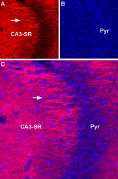 Expression of NaV1.2 in rat hippocampusImmunohistochemical staining of rat hippocampus using Anti-SCN2A (NaV1.2)-ATTO Fluor-594 Antibody (#ASC-002-AR). A. Staining of NaV1.2 (red) appears in the stratum radiatum (CA3-SR) apical dendrites (arrow) of pyramidal neurons (Pyr). B. DAPI is used as the counterstain (blue). C. Merge images of A and B.
- Wu, L. et al. (2002) NeuroReport 13, 2547.
- Fang, X. et al. (2002) J. Neurosci. 22, 7425.
- Fjell, J. et al. (2000) NeuroReport 11, 199.
- Baker, M.D. and Wood, J.N. (2001) Trends Pharmacol. Sci. 22, 27.
- Lai, J. et al. (2003) Curr. Opin. Neurobiol. 13, 291.
- Isom, L.L. (2001) Neuroscientist 7, 42.
- Catterall, W.A. et al. (2003) Pharmacol. Rev. 55, 575.
- Catterall, W.A. et al. (2008) J. Neurosci. 28, 11768.
Voltage-gated sodium channels (NaV) are essential for the generation of action potentials and for cell excitability.1 NaV channels are activated in response to depolarization and selectively allow flow of Na+ ions. To date, nine NaV α subunits have been cloned and named NaV1.1-NaV1.9.4-5 The NaV channels are classified into two groups according to their sensitivity to Tetrodotoxin (TTX): TTX-sensitive (NaV1.1, NaV1.2, NaV1.3, NaV1.4, NaV1.6 and NaV1.7) and TTX-resistant (NaV1.5, NaV1.8 and NaV1.9).2-3
Mammalian sodium channels are heterotrimers, composed of a central, pore-forming α subunit and two auxiliary β subunits. The expression of the α subunit isoform is developmentally regulated and tissue specific. Sodium channels in the adult central nervous system and heart contain β1 through β4 subunits, whereas sodium channels in adult skeletal muscle have only the β1 subunit.6,7
NaV1.2, also known as SCN2A, is primarily expressed in the central nervous system (CNS) and is localized in unmyelinated or premyelinated axons and dendrites. Mutations in the NaV1.2 channel have been identified in different types of epilepsy.
Anti-SCN2A (NaV1.2) Antibody (#ASC-002) is a highly specific antibody directed against an epitope of the rat protein. The antibody can be used in western blot, immunoprecipitation, immunohistochemistry, and immunocytochemistry applications. It has been designed to recognize NaV1.2 from rat, human, and mouse samples.
Anti-SCN2A (NaV1.2)-ATTO Fluor-594 Antibody (#ASC-002-AR) is directly labeled with an ATTO-594 fluorescent dye. ATTO dyes are characterized by strong absorption (high extinction coefficient), high fluorescence quantum yield, and high photo-stability. The ATTO-594 fluorescent label belongs to the class of Rhodamine dyes and can be used with fluorescent equipment typically optimized to detect Texas Red and Alexa-594. Anti-SCN2A (NaV1.2)-ATTO Fluor-594 Antibody is specially suited to experiments requiring simultaneous labeling of different markers.
Applications
Citations
 Powered by Bioz
- Immunohistochemical staining of mouse brain sections with #ASC-002. Tested in SCN2A-/- mice.
Planells-Cases, R. et al. (2000) Biophys. J. 78, 2878.
