Overview
- Peptide (C)DNVKYYNLARIKWD, corresponding to amino acid residues 573-586 of rat TRPC6 (Accession Q61143). 2nd extracellular loop.

 Western blot analysis of rat brain lysates:1. Anti-TRPC6 (extracellular) Antibody (#ACC-120), (1:200).
2. Anti-TRPC6 (extracellular) Antibody, preincubated with TRPC6 (extracellular) Blocking Peptide (#BLP-CC120).
- Rat kidney sections (1:50) (Ilatovskaya, D.V. et al. (2015) Sci. Rep. 5, 17637.).
 Cell surface detection of TRPC6 by indirect flow cytometry in live intact human MEG-01 megakaryocytic leukemia cells:___ Cells.
___ Cells + goat-anti-rabbit-FITC.
___ Cells + Anti-TRPC6 (extracellular) Antibody (#ACC-120), (2.5μg) + goat-anti-rabbit-FITC.
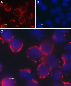 Expression of TRPC6 in rat PC12 cellsCell surface detection of TRPC6 in intact living PC12 cells. A. Extracellular staining of cells with Anti-TRPC6 (extracellular) Antibody (#ACC-120), (1:50), followed by goat anti-rabbit-AlexaFluor-594 secondary antibody (red). B. Nuclear staining with DAPI as the counterstain. C. Merged image of A and B.
- Moran, M.M. et al. (2004) Current Opin. Neurobiol. 14, 362.
- Clapham, D.E. et al. (2003) Pharmacol. Rev. 55, 591.
- Clapham, D.E. (2003) Nature 426, 517.
- Padinjat, R. and Andrews, S. (2004) J. Cell. Sci. 117, 5707.
- Huang, C.L. (2004) J. Am. Soc. Nephrol. 15, 1690.
- Winn. M.P. et al. (2005) Science 308, 1801.
- Reiser, J. et al. (2005) Nat. Genet. 37, 739.
The Transient Receptor Potential (TRP) superfamily is one of the largest ion channel families and consists of diverse groups of proteins. In mammals, about 28 genes encode the TRP ion channel subunits. The mammalian TRP superfamily comprises six subfamilies known as the TRPC (canonical), TRPV (vanilloid), TRPM (melastatin), TRPML (mucolipins), TRPP (polycystin) and the TRPA (ANKTM1) ion channels1-4.
The TRPC subfamily consists of seven proteins named TRPC1 to 7 which can be further divided into four subgroups based on their sequence homology and functional similarities: TRPC1, TRPC4, TRPC5 TRPC3, TRPC6, TRPC7 and TRPC22,5. They are highly expressed in the central nervous system and to a lesser extent in peripheral tissues.
TRPC6 can form heterotetramers with TRPC3 and TRPC7. It is primarily expressed in brain, lung and muscle. High levels of expression of the channel were also found in human platelets. Recently it was reported that TRPC6 is also expressed in the kidney where a mutated channel has been implicated in kidney failure disease6,7.
Anti-TRPC6 (extracellular) Antibody (#ACC-120) is a highly specific antibody directed against an extracellular epitope of the mouse protein. The antibody can be used in western blot, immunohistochemistry, immunocytochemistry and live cell flow cytometry applications. It has been designed to recognize TRPC6 from rat, mouse and human samples.
 Antibody")
Expression of TRPC6 in rat kidney cortex sections.Immunohistochemical staining of rat kidney sections using Anti-TRPC6 (extracellular) Antibody (#ACC-120). TRPC6 staining is detected in podocytes and darker patches are observed in hyperglycemic rat subjected to STZ treatment.Adapted from Ilatovskaya, D.V. et al. (2015) Sci. Rep. 5, 17637. with permission of Nature Publishing Group.
Applications
Citations
 Powered by Bioz
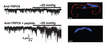 TRPC6 is required for stretch activation in myocytes.Electrophysiological recordings of myocytes incubated with Anti-TRPC6 (extracellular) Antibody (#ACC-120) in the absence (upper left) and in the presence (lower left) of the control peptide antigen. Immunocytochemical staining of living rat cerebral artery smooth muscle cells using Anti-TRPC6 (extracellular) antibody. Preincubation with the control peptide antigen blocks TRPC6 immunostaining (lower right panel).
Adapted from Gonzales, A.L. et al. with permission of the American Association for the Advancement of Science.
- Western blot analysis of MCF7 and MDA-MB-231 breast cancer cell lysates. Tested in shTRPC6-treated cells.
Diez-Bello, R. et al. (2019) Biochem. Biophys. Acta 1866, 474.
- Western blot analysis of MCF7 and MDA-MB-231 breast cancer cell lysates.
Diez-Bello, R. et al. (2019) Biochem. Biophys. Acta 1866, 474.
- Rat cerebral artery smooth muscle cells.
Gonzales, A.L. et al. (2014) Sci. Signal. 7, ra49.
- Rat kidney sections (1:50).
Ilatovskaya, D.V. et al. (2015) Sci. Rep. 5, 17637.
- Human and mouse alveolar macrophages (1:250).
Riazanski, V. et al. (2015) Proc. Natl. Acad. Sci. U.S.A. 112, E6486.
- Rat myocyte currents in electrophysiological studies.
Gonzales, A.L. et al. (2014) Sci. Signal. 7, ra49.
