Overview
- Peptide VNENTRMYIPEENHQ(C), corresponding to amino acid residues 2-15 of human CaV1.2 (exon 1B) (Accession Q13936). Intracellular, N-terminus.

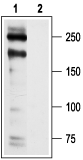 Western blot analysis of rat heart membranes:1. Anti-Human-CaV1.2 (CACNA1C) Antibody (#ACC-022), (1:200).
2. Anti-Human-CaV1.2 (CACNA1C) Antibody, preincubated with Human Cav1.2/CACNA1C Blocking Peptide (#BLP-CC022).- Mouse cortex, hippocampus, and cerebellum lysates (1:200) (White, J.A. et al. (2008) Learn. Mem. 15, 1.).
- Rat nodose ganglion neurons in culture (1:15) (Pamidimukkala, J. and Hay, M. (2004) Brain Res. 1006, 215.).
- Catterall, W.A. et al. (2003) Pharmacol. Rev. 55, 579.
- IUPHAR
All L-type calcium channels are encoded by one of the CaV1 channel genes. These channels play a major role as a Ca2+ entry pathway in skeletal, cardiac and smooth muscles as well as in neurons, endocrine cells and possibly in non-excitable cells such as hematopoetic and epithelial cells. All CaV1 channels are influenced by dihydropyridines (DHP) and are also referred to as DHP receptors.
While the CaV1.1 and CaV1.4 isoforms are expressed in restricted tissues (skeletal muscle and retina, respectively), the expression of CaV1.2 is ubiquitous and CaV1.3 channels are found in the heart, brain and pancreas. Several peptidyl toxins are described that are specific L-type channel blockers, but so far no selective blocker for one of the CaV1 isoforms have been described. These include the Mamba toxins Calcicludine (#SPC-650), Calciseptine (#C-500) and FS-2 (#F-700).
Alomone Labs is pleased to offer a highly specific antibody directed against an epitope of human CaV1.2 channel. Anti-Human CaV1.2 (CACNA1C) Antibody (#ACC-022) can be used in western blot and immunocytochemistry applications. It has been designed to recognize CaV1.2 from mouse, rat and human samples.
Applications
Citations
 Powered by Bioz
- Mouse cortex, hippocampus, and cerebellum lyastes (1:200).
White, J.A. et al. (2008) Learn. Mem. 15, 1.
- Rat nodose ganglion neurons in culture (1:15).
Pamidimukkala, J. and Hay, M. (2004) Brain Res. 1006, 215.
