Overview
- Peptide KDYPASTSQDSFEA(C), corresponding to amino acid residues 263-276 of human KV1.3 (Accession P22001). Extracellular loop between domains S1 and S2.
- Mouse brain and human Jurkat T cell lysates (1:1000).
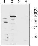 Western blot analysis of human Jurkat T cell leukemia cell lysate (lanes 1 and 3) and mouse brain lysate (lanes 2 and 4):1-2. Anti-KV1.3 (KCNA3) (extracellular)-Biotin Antibody (#APC-101-B), (1:1000).
3-4. Anti-KV1.3 (KCNA3) (extracellular)-Biotin Antibody, preincubated with Kv1.3/KCNA3 (extracellular) Blocking Peptide (#BLP-PC101).
- Mouse frozen brain sections (1:400).
- Live human Jurkat T cells (1:10).
- The control antigen is not suitable for this application.
KV1.3 belongs to the Shaker family of voltage-dependent K+ channels. The channel, encoded by KCNA3, is widely expressed in the brain, lung and osteoclasts and in several cell populations of hematopoietic origin. The prominence of KV1.3 channels in these cells (particularly in T lymphocytes) directed much research attention. It was found that KV1.3 is the main channel responsible for maintaining the resting potential in quiescent cells and regulating the Ca2+ signaling that is indispensable for normal T lymphocyte activation.1,2 Based on the central role of KV1.3 in regulating the initiation of an immune response, the channel has been recognized as a potential target for immunosuppressant drugs.1 The central role of KV1.3 in immune system cells created a real need for a specific antibody that would be able to work in flow cytometry applications.
