Overview
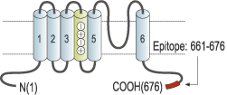
- Peptide (C)TYEQLTVPRRGPDEGS, corresponding to amino acid residues 661-676 of human KCNQ1 (Accession P51787). Intracellular, C-terminus.

 Western blot analysis of rat heart membranes:1. Anti-KCNQ1 Antibody (#APC-022), (1:200).
2. Anti-KCNQ1 Antibody, preincubated with KCNQ1 Blocking Peptide (#BLP-PC022).
- Canine left ventricle (8 μg) (Ehrlich, J.R. et al. (2004) J. Biol. Chem. 279, 1233.).
- Mouse distal colon (1:50) (Jepps, T.A. et al. (2009) Am. J. Physiol. 297, G107.).
- COS cells (Wiener, R. et al. (2008) J. Biol. Chem. 283, 5815.).
- Wang, Q. et al. (1996) Nat. Genet. 12, 17.
- Grahammer, F. et al. (2001) Gastroenterology 120, 1363.
- Mall, M. et al. (1998) Am. J. Physiol. 275, G1274.
KCNQ1 (KV7.1) is part of a voltage-gated K+ family that includes 5 other members (KCNQ1-5).
The channel has been intensively studied since it was found that together with MinK (Isk) it underlay the cardiac Iks current that controls the duration of the action potential of the human heart. Indeed mutations on either KV7.1 or MinK can be responsible for long QT syndrome, a cardiac disorder that causes arrhythmias and sudden death.
Although KV7.1 expression and function has been mainly investigated in relation with cardiac function the channel is widely distributed in epithelial tissues. There it has been implicated in physiological functions such as the regulation of acid secretion in the stomach and Cl- secretion into the colon.
Application key:
Species reactivity key:
Anti-KCNQ1 Antibody (#APC-022) is a highly specific antibody directed against an epitope of the human protein. The antibody can be used in western blot, immunoprecipitaion, immunohistochemistry and immunocytochemistry applications. It has been designed to recognize KV7.1 channel from rat, human, and mouse samples.
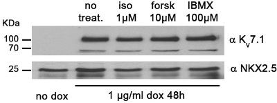
 Expression of KCNQ1 (KV7.1) in hiPSC-CMs.Western blot analysis of human induced pluripotent stem cell-derived cardiomyocytes (hiPSC-CMs) under different treatments using Anti-KCNQ1 Antibody (#APC-022). Protein levels of KCNQ1 remain the same.Adapted from Piccini, I. et al. (2017) Front. Physiol. 8, 705. with permission of Frontiers.
Applications
Citations
 Expression of KCNQ1 (KV7.1) and KCNQ5 (KV7.5) in rat smooth muscle cells.Immunocytochemical staining of rat smooth muscle cells from LCA and RCA using Anti-KCNQ1 Antibody (#APC-022) and Anti-KCNQ5 Antibody (#APC-155) shows that the expression of both channels is higher in LCA than in RCA.
Adapted from Morales-Cano, D. et al. (2015) with permission of the European Society of Cardiology.
- Guinea pig heart lysate.
Zankov, D.P. et al. (2019) Heart Rhythm 16, 108. - Rat heart lysate (1:2000).
Masuda, K. et al. (2018) J. Physiol. Sci. 68, 759. - CHO and HEK 293 transfected cells.
Meisel, E. et al. (2018) Channels 12, 89. - Human detrusor lysate.
Bientinesi, R. et al. (2017) Naunyn-Schmiedebergs Arch. Pharmacol. 390, 127. - HEK 293 transfected cells.
Moreno, C. et al. (2017) J. Mol. Cell. Cardiol. 110, 61. - Human heart tissue sample lysate (1:200).
Opthof, T. et al. (2017) Heart Rhythm 14, 265. - Human induced pluripotent stem cell-derived cardiomyocytes.
Piccini, I. et al. (2017) Front. Physiol. 8, 705. - HEK 293 transfected cells.
Inanobe, A. et al. (2015) Cell. Physiol. Biochem. 36, 1847. - Mouse transfected MDCK cells (1:1000).
Andersen, M.N. et al. (2015) Am. J. Physiol. 309, C693. - Mouse mMCD3 cell lysate (1:300).
Slaats, G.G. et al. (2015) J. Cell. Sci. 128, 4550. - MDCK cells stably expressing human KV7.1 (1:1000).
Andersen, M.N. et al. (2013) J. Biol. Chem. 288, 36841. - Xenopus oocyte membrane lysates (1:500).
Lundby, A. et al. (2013) Sci. Signal. 6, rs11.
- HEK 293 transfected cells.
Inanobe, A. et al. (2015) Cell. Physiol. Biochem. 36, 1847. - Mouse transfected MDCK cells.
Andersen, M.N. et al. (2015) Am. J. Physiol. 309, C693. - Canine left ventricle (8 μg).
Ehrlich, J.R. et al. (2004) J. Biol. Chem. 279, 1233.
- Rat rectal colon sections.
Inagaki, A. et al. (2019) Pflugers Arch. 471, 313. - Mouse cochlea sections (1:100).
Cazals, Y. et al. (2015) Nat. Commun. 6, 8780. - Rat kidney sections (1:200).
Slaats, G.G. et al. (2015) J. Cell. Sci. 128, 4550. - Guinea pig detrusor (1:200).
Anderson, U.A. et al. (2013) Br. J. Pharmacol. 169, 1290. - Mouse distal colon (1:50).
Jepps, T.A. et al. (2009) Am. J. Physiol. 297, G107.
- Guinea pig myocytes.
Zankov, D.P. et al. (2019) Heart Rhythm 16, 108. - Mouse mMCD3 cells (1:200).
Slaats, G.G. et al. (2015) J. Cell. Sci. 128, 4550. - Rat smooth muscle cells (1:100).
Morales-Cano, D. et al. (2015) Cardiovasc. Res. 106, 98. - COS cells.
Wiener, R. et al. (2008) J. Biol. Chem. 283, 5815.
- Lundby, A. et al. (2013) Sci. Signal. 6, rs11.
- Askar, S.F. et al. (2013) Cardiovasc. Res. 97, 171.
- Organ-Darling, L.E. et al. (2013) Am. J. Physiol. 304, H589.
- Alzamora, R. et al. (2010) Am. J. Physiol. 299, F1308.
- Roura-Ferrer, M. et al. (2010) J. Cell. Physiol. 225, 692.
- Roura-Ferrer, M. et al. (2008) Biochem. Biophys. Res. Com. 369, 1094.
- Xiao, L. et al (2008) Circulation 118, 983.
- Yeung, S.Y. et al. (2008) Br. J. Pharmacol. 155, 62.
- Yeung, S.Y. et al. (2007) Br. J. Pharmacol. 151, 758.
